Functions of Cells and Human Body
Content:
1. Introduction to the gastrointestinal motility
2. Motility of the stomach
3. Motility of the small intestine
4. Motility of the colon
_
Introduction to the gastrointestinal motility
The term motility is defined as involuntary mobility of human tubular organs. To ensure the efficient digestion of food is necessary not only the presence of active enzymes, but also a shift and mixing of chyme during passage through the digestive tube. For this purpose, there are two types of movements in the GIT:
1) Propulsion movements
2) Mixing movements
This division is artificial, because, in fact, both types of movements use the same mechanisms and often one can convert to the other.
推進運動
推進運動は、消化管内でチャイムを動かすための運動です。
蠕動運動

基本的な推進運動は蠕動運動と呼ばれるものです。 その原理は簡単で、ある場所の円形筋層が収縮して収縮輪を作り、それをさらに腹側に移動させるというものです。 こうして、ゆっくりと粥を押し進めるのである。 腸の膨張は、しばしば収縮輪の形成、ひいては蠕動運動の開始のきっかけとなる。 腸管が膨張すると、腸管神経系が刺激され、蠕動運動が始まる。 この刺激により、チューブの最大膨張位置から数インチ経口したところにある円形筋セグメントの収縮が引き起こされる。 蠕動運動は、ある種の化学的刺激や強い副交感神経の活性化によっても引き起こされる。 また、ある一定の間隔で自動的に発生する。 脹ら脛の経口的な収縮に加え、脹ら脛の経口的な受容性弛緩と呼ばれるものが起こる。 これは、弛緩した管が流れる際の抵抗を少なくするためで、この弛緩によりチュームの移動が容易になる。
口腔方向にも推進運動が起こるが、数ミリで消失してしまう。
混合運動
混合運動は、栄養的に重要な成分の全量を酵素にさらし、腸の内壁に接触させて吸収させるために、チャイムを常に混合することを保証します。
分節
分節は、よく理解された混合運動です。 円形平滑筋の数センチ離れた部分が繰り返し収縮するようなイメージです。 収縮した部分は、1回の分節化で異なる。 このように、分節化された胆汁は、再び分割され、半分が前の胆汁の一部と結合し、もう半分が次の胆汁の一部と結合する。 2つの外側の部分の半分が常に結合する場所を失い、新しい部分を形成するため、部分の数は徐々に増加し、一方その量は減少する。
_
胃の運動
胃はその筋肉の配列のおかげで、三つの機能をうまくこなしているのである。
1) 食物の混合
2) 多量の食物の貯蔵
3) 十二指腸へ排出
食物の混合
胃にチャイムが存在するまでのことである。 その上3分の1に弱い収縮波(混合波と呼ばれる)が形成される。 これらは20秒ごとに規則的に現れ、平滑筋の自動性に基づいている。 混合波が胃の本体から前門部まで伸びると、より強力になり、チムを幽門部まで強く押し出すようになる。 しかし、幽門括約筋が閉じられると、盲端と混合のみ、それぞれ高圧下で収縮環の動きに逆らって逆流するようにチュームが遭遇する。 この現象をretropulsionと呼ぶ。
pyloric sphincterは決して完全に閉じていないことに注意すると良いだろう。 しかし、十二指腸がその組成を検査し、それに応じて胃の運動量を調整するという重要な機能をもっている。
胃排出
胃排出も逆流と同じメカニズムである。 しかし、同時に、胃液が幽門を通過する際の抵抗が減少し、幽門括約筋の弛緩が起こります。 このため、その時々の幽門の抵抗力に応じて、一定量の胃液が十二指腸に流れ込むことができる。
幽門括約筋は、通常の円形の筋肉をさらに厚くしたようなものです。 彼は胃の残りの部分と比較して、幽門の約2倍の厚さを持っています。 通常、その調子は、抵抗が液体の通過のために十分に小さいが、固体チュームの通過のために大きすぎるように設定されています。 胃液は何度も逆流混合を繰り返し、胃液と十分に混ざり合って十二指腸に進む必要がある。
胃排出は、2つのグループに分けられるいくつかの要因によって制御されています。
1) 胃要因
2) 十二指腸要因
胃要因
胃要因は通常、混合波の周波数を上げるか幽門緊張を低下させて胃排出量を増加させる。 胃の中に大量の食物(特に肉などのタンパク質を多く含む食物)がある場合、活性化される。 ペプチドが肛門粘膜に接触すると、消化管ホルモンであるガストリンが分泌される。
Gastrin has the following effects:
1) Increases the production of gastric juice that has low pH
2) Increases the frequency of spontaneous motor activity of the stomach (mixing waves)
3) Decreases the pyloric sphincter tone
Note that if there is a sudden increase of the frequency of mixing waves and a decrease of pyloric sphincter tone, increased efficiency of the pyloric pump occurs. This is the main mechanism of increased gastric emptying.
Duodenal factors
These are mostly inhibitory signals that block gastric emptying. There are two main groups:
1) Nerve feedback to enterogastric system
2) Feedback control through the gastrointestinal hormones
Nerve feedback to enterogastric system
If large volume of chyme passes through the pyloric sphincter into the duodenum, there is a distension of its wall leading to a reflex that slows down or completely stops gastric emptying. This signal is mediated:
1) Directly by the enterogastric system
2) Through the paravertebral sympathetic ganglion
3) Through the vagus nerve to the brainstem and back
All of them are called pyloric reflexes. Their effect is dual:
1) Decreased frequency of mixing waves
2) Increased tone of the pyloric sphincter
This slows down the mechanism of the pyloric pump.
In addition to the volume of the chyme, the pyloric reflexes are activated by the low pH (3.5-4)、チャイム中のペプチドの高濃度、高張性・低張性によって活性化する。
消化管ホルモンによるフィードバック制御
十二指腸の上皮細胞は特定の種類の栄養素に対して感覚的な活動を示しています。 pH、ペプチド濃度、張力が変化すると、幽門反射を起こします。
CCK – コレシストキニン
このホルモンには、3つの重要な作用があります。
セクレチン
セクレチンは、胃液の低いpHへの応答として十二指腸細胞によって産生されます。 It inhibits gastric emptying.
GIP – gastric inhibitory peptide
GIP is produced as a response to the high lipid content in the chyme. Although it has an inhibitory effects on the gastric motility and especially on the the pyloric pump it is the weakest one of all three hormones.
_
Motility of the small intestine
Contractions of the muscle layers of the small intestine can be divided into two groups:
1) Segmentation contractions
2) Propulsion contractions
Segmentation contractions
Process of the segmentation has already been discussed above. We only briefly discuss its causes here. Segmentation is a manifestation of electrical slow-waves, which represent action potentials generated by the automaticity of smooth muscle. Maximal frequency of these slow waves is 12/min. したがって、分節化も1分間に12回まで起こる可能性がありますが、非常にまれなケースに限られます。
推進収縮
そのメカニズムについては、導入部ですでに説明したとおりです。 小腸の収縮輪は約0.5〜2cm/minの速度を持っています。 近位部では速く、遠位部では遅くなる。 1つの収縮環は最大で10cm移動し、その後、外に出て、新たな収縮環を待ちます。
小腸の運動制御
小腸では毎食後、推進運動が盛んになる。 これは、小腸にチャイムが存在することと、胃腸反射の両方によるものである。 この反射は胃壁の伸張に対する反応であり、腸の運動性を高める。 この反射の構成要素は、完全に腸管神経叢にあります。 さらに、CCK、ガストリン、インスリン、モチリンといったホルモンが作用します。 これらのホルモンは食後に分泌され、推進運動と混合運動の頻度を増加させる。 逆にセクレチンやグルカゴンは小腸の運動を抑制する。
回盲弁
この弁の機能は、大腸から小腸へのチャームの還流を防ぐことである。 実際には弁ではなく、盲腸に突き出た回腸末端の開口部である。 しかし、筋肉の量が多いため、弁として機能する。 その機能は抵抗に依存する。
_
大腸の運動
大腸は主に電解質と水を吸収する機能と固形廃棄物を体外に排出する前に貯蔵する機能の2つを担っています。 この2つの機能には、大規模な運動は必要ありません。 したがって、縦筋層はtaeniaに削減結腸にある。 これらは、大腸の全長に沿って伸びる3本の帯状の筋肉を表している。
腸管運動
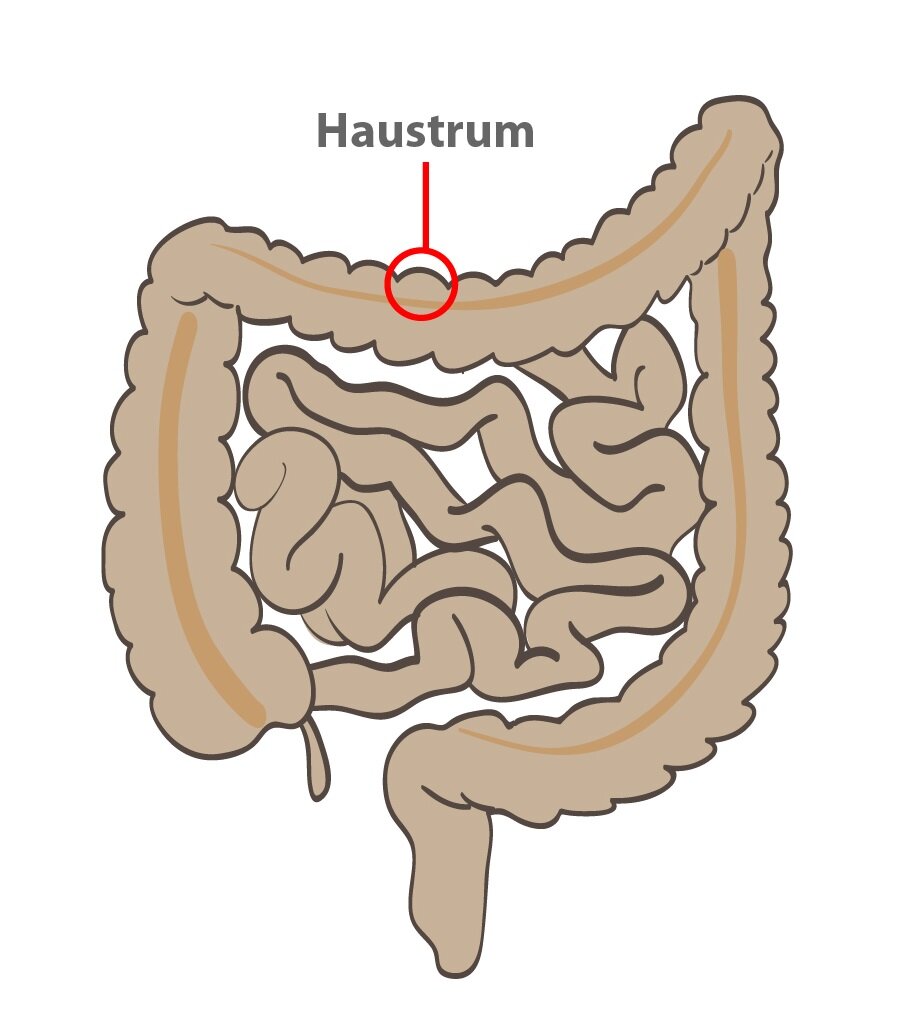
 大腸の混合運動(改良型分節運動)である。 まず、円筋の収縮がある。 その後、毛細血管が収縮してハウストラムを形成する。 ハウストラムは、比較的大きな膨らみが連続する大腸の特徴的な外観を形成する。 大腸が収縮している間、ハウストラの内部は圧力が上昇する。 約30秒後に圧力は最大になり、次の60秒後には胞体が消えます。
推進運動
推進運動の大部分は、盲腸からS状結腸へ徐々に進行する噴門によって決定されます。
しかし、もっと速い推進運動もあり、それは1日3回、食後1時間程度、15分間だけ行われます。 それは蠕動運動を連想させる。 収縮輪というものがあり、これが徐々に腹腔方向へ移動する。 横行結腸に発生し、約15分間は正常な排便活動が消失する。
結腸運動の調節
記述された結腸運動はすべて、二つの反射が引き起こされたときに強度と頻度が増加する。
1) Gastrocolic reflex
2) Duodenocolic reflex
Gastrocolic reflex
Gastrocolic reflex is triggered by a high tension in the stomach wall. Myenteric plexus transports this signal through to the colon that increases the frequency of haustra formation.
Duodenocolic reflex
Duodenocolic reflex is triggered by a high tension in the duodenal wall. Signal spreads through the myenteric plexus to the colon and increases the frequency of action potentials in the smooth muscle cells. That increases speed of the propulsion movements.
Subchapter Author: Patrik Maďa
![]()