Spine Anatomy
Anatomy & Function
The spine is remarkable and complex, and a basic understanding of its anatomy and function is especially important for patients managing spinal disorders.
Spine Functions
The three main functions of the spine include:
- Protecting the spinal cord, nerve roots & several of the body’s internal organs
- Providing structural support & balance to maintain an upright posture
- Enabling flexible motion of the spine & body
Regions of the Spine
The spine is divided into four main regions: cervical, thoracic, lumbar and sacral. Each region has specific characteristics and functions.
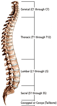
Cervical Spine
The neck region of the spine is known as the cervical spine. 上から順にC1~C7と略される7つの椎骨で構成されています。
第1頸椎(C1)はアトラスと呼ばれ、脳幹と脊髄を保護し、頭蓋骨を支え、頭を大きく動かすことができる。 アトラスはリング状になっていて、頭蓋骨を支えている。 C2は軸と呼ばれます。 C2は円形で、アトラスの輪の中に上向きに突き出た鈍い釘のような構造(歯状突起または「デンス」と呼ばれる)を持っています。 アトラスと軸が一緒になって、頭の回転と旋回を可能にしています。
胸椎
最後の頸椎の下には、胸椎の12個の椎骨があります。 上から順にT1~T12と略される。 T1が一番小さく、T12が一番大きい胸椎です。
棘突起が長いことに加え、肋骨の付着が胸椎の強度を高めています。
棘突起が長いことに加え、肋骨の付着が胸椎の強度を高めている。これらの構造により、胸椎は頸部や腰部よりも安定している。
腰椎
腰椎にはL1~L5(最大)の5つの椎骨がある。 各腰椎の大きさや形は、体のほとんどの重さを支えるように設計されています。
腰椎の各構造要素は、頸椎や胸椎の同様の構成要素よりも大きく、幅が広く、広い。
腰椎の可動域は胸椎よりも広いが、頸椎よりは狭い。
腰椎のファセット・ジョイントは、回転を制限しつつ、大きな屈曲と伸展の動きを可能にする。
仙骨は、骨盤の後ろに位置する。 5つの骨(S1~S5と略す)が三角形に融合し、仙骨を形成しています。 仙骨は、背骨と骨盤をつなぐ2つの腰骨の間に収まっています。 最後の腰椎(L5)は、仙骨と関節(動く)しています。
骨盤 & 頭蓋骨
一般的に脊椎の一部とは見なされていませんが、骨盤と頭蓋骨は脊椎と密接に関連し、人のバランスに大きな影響を与える解剖学的構造物です。
脊椎平面
解剖学をさらに理解し説明するために、脊椎専門家はしばしば特定の身体平面(body planes)について言及します。 ボディ・プレーンとは、特定の解剖学的領域を定義するために使用される、想像上の平らな二次元表面である。
表 1
| 用語 | Meaning |
| Meaning |
Spinal Curves
When viewed from the front (coronal plane), the healthy spine is straight. A sideways curve in the spine is known as scoliosis. When viewed from the side (sagittal plane) the mature spine has four distinct curves. These curves are described as being either kyphotic or lordotic.
A kyphotic curve is a convex curve in the spine (i.e. convexity toward the back of the spine).
椎骨の構造
すべての椎骨は、最初の2つの頸椎を除いて、同じ基本要素で構成されています。 椎骨の外殻は皮質骨でできています。 このタイプの骨は密度が高く、しっかりしていて丈夫です。 椎骨の内部には海綿骨があり、皮質骨より弱く、蜂の巣のような緩やかな構造をしています。
椎骨は、以下の共通要素で構成されています。
- 椎体。 椎骨の中で最も大きな部分。 上から見ると、やや楕円形の形をしている。 横から見ると砂時計のような形をしており、両端が太く、真ん中が細くなっている。 椎体は強固な皮質骨で覆われ、海綿骨で満たされている。
- 柱。 強靭な皮質骨でできた2本の短い突起で、椎体の背面から突き出ている。
- 突起:比較的平らな2枚の骨の板で、左右の小柱から伸び、正中線で結合している。 関節突起、横突起、棘突起の3種類がある。 靭帯や腱の接続点として機能する。 4つの関節突起は、隣接する椎骨の関節突起と結合し、小顔の関節を形成する。 小面体関節と椎間板の組み合わせにより、脊椎の運動が可能になる。 棘突起は、2つの薄板が結合する部分から後方に伸びており、椎骨を動かすためのレバーとして機能する。 横突起は、各椎骨の左右に突き出ている小さな骨片です。 これらの突起は、筋肉や靭帯の付着部として機能し、胸椎の肋骨の関節点として働く。 各椎体の上部(上側)と下部(下側)は、内板で「覆われて」いる。 内板は椎間板と一体化した複雑な構造をしており、椎間板を支えるのに役立っている。 小柱の上面には小さな切り欠きがあり、下面には深い切り欠きがある。 椎骨を積み重ねると、この台座の切り欠きが椎間孔と呼ばれる部分を形成する。
顔面関節
脊柱の関節は、椎体の後方(背中側)に位置しています。 これらの関節は、背骨がさまざまな方向に曲がり、ねじれ、伸びるのを助ける。 関節は動きを可能にしますが、過伸展や過屈曲(つまりむち打ち症)のような過度の動きも制限します。
各脊椎骨には2つの小面体関節があります。
体の他の関節と同様に、各関節は結合組織のカプセルに包まれており、関節に栄養と潤滑を与える滑液を分泌しています。
椎間板
椎体の間には、椎間板と呼ばれる「クッション」のようなものがあります。 それぞれの椎間板は、運動時のストレスや衝撃を吸収し、椎骨同士がぶつかり合うのを防いでいます。 椎間板は、血管が通っていない体の中で最も大きな構造物です。
各ディスクは、環状線維と髄核の 2 つの部分から構成されています。
環状線維
タイヤのように頑丈な構造で、ゲル状の中心部である髄核を包んでいます。
環状骨は、水と丈夫な弾性コラーゲン繊維の層で構成されています。
環状部は、水と丈夫な弾性コラーゲン繊維の層からなり、繊維はラジアルタイヤの構造に似て、異なる水平角度に配向しています。
このように、椎間板の中心部は、ゲル状の弾性物質で満たされています。
椎間板の中心部はゲル状の弾性体で満たされており、線維輪とともに椎骨から椎骨への応力や体重の伝達を担っています。 髄核は、環状線維と同様に、水、コラーゲン、プロテオグリカンから構成されています。 ただし、髄核はこれらの物質の割合が異なります。
脊髄と神経根
脊髄は、小指の幅ほどの細長い円筒形の構造をしています。 脊髄は脳幹のすぐ下から始まり、第一腰椎(L1)まで伸びている。 その後、脊髄は馬尾に似た神経の集まりである馬尾となる円錐髄膜と混ざり合う。 脊髄神経根は、運動と感覚を刺激する役割を担っている。
脳と脊髄は中枢神経系(CNS)を構成する。 The nerve roots that exit the spinal cord/spinal canal branch out into the body to form the peripheral nervous system (PNS).
Between the front and back portions of the vertebra (i.e. the mid-region) is the spinal canal housing the spinal cord and the intervertebral foramen. The foramen are small openings formed between each vertebra. These “holes” provide space for the nerve roots to exit the spinal canal and further branch out to form the peripheral nervous system.
Table 2
| Type of Neural Structure | Role/Function |
| Brain Stem | Connects the spinal cord to other parts of the brain. |
| Spinal Cord | Carries nerve impulses between the brain and spinal nerves. |
| Cervical Nerves (8 pairs) | These nerves supply the head, neck, shoulders, arms, and hands. |
| Thoracic Nerves (12 pairs) | Connects portions of the upper abdomen and muscles in the back and chest areas. |
| Lumbar Nerves (5 pairs) | Feeds the lower back and legs. |
| Sacral Nerves (5 pairs) | Supplies the buttocks, legs, feet, anal and genital areas of the body. |
| Dermatomes | Areas on the skin surface supplied by nerve fibers from one spinal root. |
Table 3
| Ligament Name | Description |
| Anterior Longitudinal Ligament (ALL) A primary spine stabilizer |
About 1 inch wide, the ALL runs the entire length of the spine from the base of the skull to the sacrum. It connects the front (anterior) of the vertebral body to the front of the annulus fibrosis. |
| Posterior Longitudinal Ligament (PLL) A primary spine stabilizer |
About 1 inch wide, the PLL runs the entire length of the spine from the base of the skull to sacrum. It connects the back (posterior) of the vertebral body to the back of the annulus fibrosis. |
| Supraspinous Ligament | This ligament attaches the tip of each spinous process to the other. |
| Interspinous Ligament | This thin ligament attaches to another ligament, called the ligamentum flavum, that runs deep into the spinal column. |
| Ligamentum Flavum The strongest ligament |
This yellow ligament is the strongest one. It runs from the base of the skull to the pelvis, in front of and behind the lamina, protecting the spinal cord and nerves. |
筋肉と腱
脊椎の筋肉システムは複雑で、いくつかの異なる筋肉が重要な役割を担っています。 筋肉の主な機能は、背骨を支え安定させることです。 特定の筋肉は、解剖学の特定の部分の動きと関連している。 例えば、胸鎖乳突筋は頭部の動きを補助し、大腰筋は大腿部の屈曲に関連する。
筋肉は、個々またはグループで、筋膜によってサポートされている。 筋膜は、強い結合組織です。 筋肉を骨に付着させる腱は、筋膜の一部です。
脊椎の筋肉は、屈筋、回旋筋、伸筋と呼ばれます。
脊椎についてもっと知る
解剖学と機能についてもっと知るには、以下の評判の良い資料を活用してください:
- Spine-Health.com
- American Academy of Orthopedic Surgeons
Princeton Brain & Spine knows that many of our patients like to research their condition and treatment
and look for useful resources on the internet. However, many find this a frustrating experience.
Princeton Brain & Spine is pleased to offer you a unique health and wellness search service for internet
information that:
- Allows you to be an intelligent efficient user of the internet for health information
- Allows you to
find resources for top health and wellness sites in one place that are relevant to your
questions - Allows you to find resources in formats that you can “digest” and use to learn