Trendelenburg’s Test and Trendelenburg’s Gait
Introduction
The aim of the Trendelenburg’s test is to assess the strength of the hip abductors, specifically the gluteus medius and minimus.1
Background
The test is named after Friedrich Trendelenburg, who described the test in 1895.
Hip abductors assessed in Trendelenburg’s test 2
The hip abductors which are assessed with Trendelenburg’s test are shown in the table below.
| Origin | Insertion | Action | Nerve | |
| Gluteus medius | Gluteal surface of the ilium | Greater trochanter of the femur | All fibres: abduct the hip Anterior fibres: flex and medially rotate the hip Posterior fibres: extend and laterally rotate the hip |
Superior gluteal nerve |
| Gluteus minimus | Gluteal surface of the ilium | Greater trochanter of the femur | Abduct, medially rotate and flex the hip | Superior gluteal nerve |
Gait cycle in brief
The gait cycle is the sequence of events between the time one foot touches the ground and the time the same foot returns to the same position. Events of the gait cycle include heel strike; foot flat; push off (heel off + toe-off) and the stance and swing phases.1
During the foot flat phase of the gait cycle, the contralateral leg is lifted to do the heel strike. 対側の脚が空中にあり、同側の脚が床にある間(ミッドスタンス/シングルレッグスタンス)、この段階での股関節の位置は、股関節外転筋の強さを評価する際に重要です(体のすべての重量を片方の脚で負担しているため)。
股関節外転筋が十分に強くない場合、または痛みの抑制があると、患者は上体の重量を患肢に分散させることができません。

Trendelenburgテストはいつ行うのですか?
患者が足を引きずったり、股関節の痛みを訴えたりしたとき、または歩行を評価する通常の身体検査の一環として行います。
トレンデレンブルグ試験の実施方法
詳しくは股関節検査ガイドを参照してください。
患者の位置
患者が一人で立っていてもよい場合は、患者の後ろに立ってください。
片足立ち
患者さんに両足を順番に地面から上げてもらいます。
患者さんが右足を上げると、左股関節外転筋がテストされます。
患者さんが左足を上げると、右股関節外転筋がテストされます。
観察
患者の腸骨稜に手を置きます(これを行う前に、各ステップを明確に説明し、同意を得るようにしてください)
サポートされていない側の股関節が持ち上がるか下垂するかを注意深く観察します。
患者があなたの腕をサポートとして使用している場合、その股関節外転筋が彼らの体重をサポートできない場合は、あなたの一方の腕に押されると感じます。
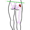
内転筋は、片足立ちのときに骨盤を水平に保つのに十分な強さが必要ですが、支えていない部位で骨盤がわずかに上がるのは正常です(これは内転筋の収縮によるものです-図1参照)。3
陽性徴候
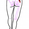
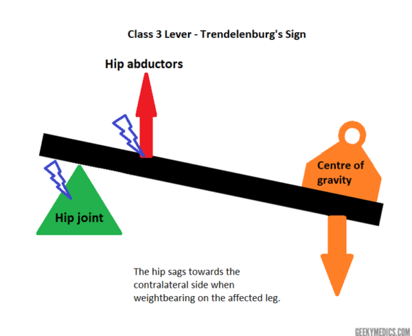
トレンデレンブルグ陽性徴候(病的)には、外転筋が体重を支える大腿骨に向かって股関節を安定させられないために、支えていない側の骨盤がたるんでいることが含まれます(図2参照)。
Trendelenburg’s gait
Trendelenburg’s gait involves excessive up-down motion of the pelvis whilst walking.
It occurs as a result of compensatory mechanisms due to the drooping pelvis.3
Unilateral positive Trendelenburg’s sign produces a lurching gait.
Bilaterally positive Trendelenburg’s sign produces a waddling gait.
Pathophysiology
Imagine a chair…when you remove a leg what happens? The chair will normally fall towards the side that had the leg removed. The human body is the same, the centre of gravity passes midway through the body and through the pubic symphysis.
When standing on one leg, the centre of gravity, much like the chair, shifts to the unsupported leg.
私たちが倒れないのは、股関節外転筋が骨盤を体重のかかっている方の足の大腿骨の方に引っ張っているからです。
股関節のテコの仕組み
ほとんどの骨格筋の動作にはテコ、つまりテコを使って物を動かすことが関わっています。
てことは、力を加えると支点と呼ばれる固定点上を動く剛体の棒です。
人体では、関節が支点となり、骨がてこの役割を果たします。
力は、荷重と努力という形で、てこにわたって発揮することが可能です。
筋肉の収縮は、骨上の筋肉の挿入ポイントで適用される力を提供します。
負荷は、骨そのものと、それを覆う組織です。
レバーには3種類あり、支点、負荷、力が互いにどのように配置されるかによって異なります6。
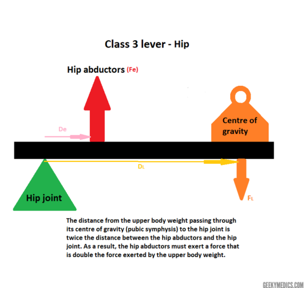
この場合、股関節はクラス3のレバーです。
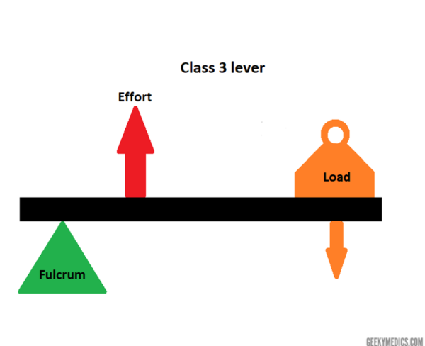
股関節のクラス3は、力(股関節外転筋が及ぼす力)がレバー(大腿骨の首)をはさんで支点(股関節)と負荷(上体の重さ)の間に位置づけられること意味しています。 Class 3のレバー
構造物を絶対に水平に保つためには、支点からの距離にわたってかかる努力と荷重の力が釣り合って、互いに打ち消し合う必要があります(図4)。
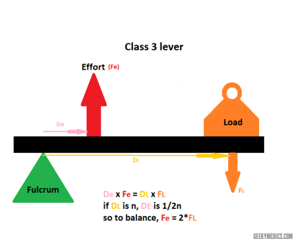
これを股関節に適用すると(図5)、股関節と股関節外転筋の間の距離は股関節と重心(恥骨結合)の間の距離の半分なので、股関節外転筋が発揮する力は上体重量が発揮する力の倍でなければならないということがわかります。
トレンデレンブルグ徴候の原因
股関節に影響するあらゆる問題は、正のトレンデレンブルグ徴候(図6)を引き起こす可能性があります。 上半身の体重を変えたり、影響を与えることはできないため、股関節や大腿骨(骨格の異常)、股関節外転筋(神経筋の異常)に影響を与える状態が残る。8
Skeletal abnormalities
トレンデレンブルグの徴候をもたらすことができる骨格異常の例である。7,8,9
- 股関節の発達異形成(DDH)-股関節の自然脱臼を引き起こす疾患のスペクトル
- 大腿骨頚部と大腿骨軸の角度が120°未満
- Slipped capital femoral epiphysis (SCFE) -大腿骨骨幹部が骨端に対してスライドする
- Legger-Calve->
- Legg-Calve->
- Developmental Disease Of The Hipthies -股関節の自然脱落を引き起こす障害のスペクトル
- Developmental Degree -大腿骨頸部と骨端間の角度。ペルテス病 – 大腿骨頭壊死を特徴とする小児股関節疾患
- 慢性亜脱臼/脱臼 – 股関節の不安定性をもたらす
- 大転子骨折(急性または非急性)liunited) – 大殿筋と小殿筋の挿入が損なわれる
神経筋の異常
トレンデレンブルグの兆候をもたらす可能性がある神経筋の異常の例。7,8,9
- 上臀部神経損傷-股関節全置換術/半関節形成術に伴う、または外傷によるものが多い
- L5神経障害-上臀部神経はL4、L5、S1脊髄根から形成されており、しばしば足の低下を伴う
- 中臀筋および小臀筋の弱化または損傷(e.例:断裂または腱炎)
- 脊髄炎-股関節外転筋の麻痺を引き起こす
- 筋ジストロフィーおよびその他の神経筋障害
- 臀部の弱さを引き起こす痛みの抑制(例:。 変形性股関節症)
検査の限界
トレンデレンブルグ徴候の陽性は、骨壊死の初期段階で隠されることがあり、骨盤の落ち込みは、股関節外転機構は正常だが機能が不十分な健常者に生じることがあります8。
偽陽性は、理解/遵守の欠如、重大な股関節痛、および脊柱側弯症 (肋骨縁が腸骨稜に衝突することがある) で起こることがあります。
患者が体重を支える股関節上に体幹 (と重心) を移動して補正すると偽陰性が起こることがあるので、上体の動きに注意します 9
さらなる検査
目標は異常歩行の根本原因を特定することにあります。
画像診断:
- 局所関節のX線(例:膝、股関節、腰椎)
- 超音波、CTまたはMRIを検討する
臨床検査:
- ベースライン血液検査は全身疾患の証拠をスクリーニングするために行うことがある。
特別な検査:
- 筋生検
- 神経伝導検査
鑑別診断
トレンデレンブルグ歩行と他のよくある歩行パターン(3、4)を区別できることは有用です
- 無痛歩行(antalgic gait)。 体重負荷時の痛みにより発生し、痛みのある側を短くしか踏まない
- 短足歩行:両足の長さの違いにより発生し、短い側の骨盤の一貫した傾きが観察される
キーポイント
- Trendelenburgのテストでは股関節外転筋の強さを評価しています。
- ルーチンの歩行検査の一部として、または患者が足を引きずったり股関節の痛みを訴えたりしたときに行われる。
- トレンデレンブルグ徴候が陽性(病的)だと、外転筋が体重を支える大腿骨に向かって股関節を安定させることができず、支えていない側の骨盤にたるみが生じている。
- If a positive sign is present, Trendelenburg’s gait may also be observed.
- Causes of a positive Trendelenburg’s sign include skeletal and neuromuscular abnormalities affecting the hip and hip abductors respectively.
- Trendelenburg’s gait should be differentiated from antalgic gait (due to pain) and short-leg gait (due to differences in leg length).
Reviewer
Miss Margaret Brooks
Orthopaedic Registrar
University Hospitals of Birmingham NHS Foundation Trust
- The Orthopaedic Physical Examination 2nd ed, Elsevier, 2005
- Trail Guide to the Body 3rd ed, Books of Discovery, 2005
- Hamblen and Simpson. Adams’s Outline of Orthopaedics 14th ed, Churchill Livingstone Elsevier, 2010
- Solomon, Warwick and Nayagam. Apley and Solomon’s Concise System of Orthopaedics and Trauma, 4th ed, Taylor & Francis Group, 2014
- Duckworth and Blundell. Lecture Notes: Orthopaedics and Fractures 4th ed, Wiley-Blackwell, 2010
- Marieb and Hoehn. Human Anatomy and Physiology, 7th ed, Benjamin Cummings, 2011
- Azar, Beaty and Canale. Campbell’s Operative Orthopaedics 13th ed, Elsevier, .2017
- Gandbhir VN, Rayi A. Trendelenburg Gait, StatPearls Publishing; 2020 Jan –
- Hardcastle and Nade. The Significance of the Trendelenburg Test (JBJS Br, 67-B no 5. 1985)
Images
- Figure 1 and 2: Illustrated by Leyla Noury, Sketchbook, 2020
- Figures 3 to 6: Illustrated by Leyla Noury, Paint, 2020